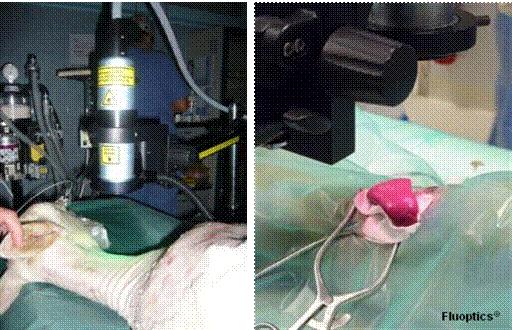
Fluoptics – Mobile in vivo imaging cameras for large and small animals, oncology research, cardiovascular research, drug tracking and therapeutic evaluation. Fluoptic offers more than just an optical imaging system. Numerous optional near-infrared fluorescent probes help you delve deeper into the development of the disease until it helps you come up with a reasonable solution. The left picture shows the carotid jugular vein imaging of the ewe, and the right picture shows the separation of fluorescently labeled tissues in the spleen of rhesus monkeys. Fluobeam® Imaging System Features: Cardiovascular Research---In the thrombus model, as well as other cardiovascular animal models, observe surgery, drug treatment effects, objective and dynamic evaluation of treatment options (for example, in vascular grafting, bypass surgery, rapid and rapid evaluation of surgical success) Whether or not). Lymph node image---The lymph node is an important immune organ of the human body. It is closely related to many major diseases such as tumors, inflammation, etc. The imaging of lymph nodes helps us to better grasp the development of these diseases. In vivo distribution of drug-loading systems/fluorescent substances, metabolic studies--New drug delivery systems such as nanoparticles/liposomes are modified by specific targeting groups to achieve active targeting functions, such as RGD tumor targeting, Brain targeting of ferritin and so on. Clinical application: 3D Folding Face Mask,3D Folding Protective Mask,3D Foldable Ffp2 Respirator,3D Folding 5 Layer Disposable Respirator Henan Aklly Filter Engineering Co., Ltd , https://www.akllyfilter.com
With the rapid development of medical and biological research, researchers are increasingly hoping to directly monitor the cellular activities and gene expression in living organisms, and effectively study the physiological processes of transgenic animals, such as tumor growth and metastasis, infection in living animals. Sexual disease development process, etc. As an emerging imaging technology, living animal optical imaging technology has become an ideal method for imaging of living animals because of its simple operation, intuitive results, high sensitivity and low cost.
In vivo optical imaging of living animals is divided into bioluminescence and fluorescence. Fluorescence imaging is increasingly favored by researchers because of its low cost, strong signal and simple operation. However, there are various drawbacks in traditional fluorescence imaging applications, such as autofluorescence interference of animal tissues and tissue characteristics of light. Absorption, etc. all affect the application of traditional fluorescence imaging.
Since the excitation light generated by the near-infrared laser has deeper tissue penetration than white light, even deeper, smaller targets can be detected. Therefore, near-infrared dyes are particularly suitable for in vivo fluorescence imaging (in vivo imaging), an emerging field that has rapidly developed in recent years. Since the autofluorescence of cells and tissues is minimal in the near-infrared region, near-infrared dyes provide higher specificity and sensitivity when detecting complex biological systems.
The Fluobeam series of in vivo imaging systems developed by Fluoptic overcomes the shortcomings of traditional fluorescent in vivo imaging, using near-infrared dye labeling and real-time imaging to provide researchers with more accurate and sensitive experimental data, and qualitative and quantitative research.
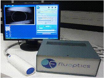
Because Fluoptic is an open working environment, it is not limited by the size of the imaging box. It can complete the living image of small animals. It is also suitable for large animal imaging. New Zealand rabbits, rhesus monkeys, even sheep and orangutans can be completed by one system. Eliminate the trouble of buying different instruments for different animals, economical, easy to operate, and save space.
Flexible and portable;
Perfect imaging is possible without the need for a darkroom;
Real-time monitoring, data can be output in multiple formats in pictures, vido;
Suitable for all fluorescent probes above CY5.
Fluobeam® application features:
Tumor Research---Using fluorescent probes to detect tumor occurrence, development, and lesion metastasis, and provide qualitative and quantitative research results.
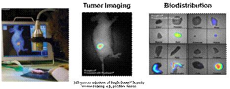

Tumor resection---- Combined with tumor-specific fluorescent probes, it can distinguish normal tissues and lesions early and clearly, providing scientific basis for accurate tumor resection; especially for patients with large tumor metastasis, traditional surgical resection If the area is too large to be implemented, and how the tiny lesions are completely removed after the lymphatic metastasis of the tumor occurs.
Interventional treatment----in the heart bypass, vascular graft surgery, the success of the operation, can be based on the accumulation of fluorescent substances or not, to determine the surgical results in a timely manner.