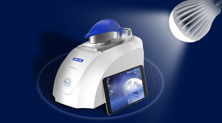
A spectrophotometer is an instrument that quantitatively and qualitatively analyzes a substance by spectrophotometry. Since the quantitative sample size of nucleic acid, protein quantification and bacterial growth concentration is small, an ultra-micro spectrophotometer has emerged. In recent years, ultra-micro spectrophotometers have replaced ordinary spectrophotometers as the new darling of molecular biology laboratories, and are widely used in the fields of proteomics and genomics in life science laboratories. Compared with traditional spectrophotometers, the ultra-micro spectrophotometer has the unique advantages of the former: (1) The required sample volume is small, only 0.5-2 μl; (2) No need for cuvette, use the pipette to directly drop the sample onto the detection platform. When measuring, the sample will automatically form a liquid column. After the test is completed, just clean the sample from the detection platform with clean absorbent paper. Yes, avoiding the cross-contamination caused by the cleaning of the cuvette; (3) Generally has multiple optical paths (automatic selection of motor control) [Automatic adjustment of optical path 1.0 mm, 0.2 mm, 0.1 mm and 0.05 mm in BIO-DL MicroDrop base mode] , while the optical path of traditional spectrophotometer is The 10 mm, ultra-micro spectrophotometer sample does not need to be diluted, and the measurement range can reach 50 times that of the conventional spectrophotometer [dDNA detection range of BIO-DL MicroDrop pedestal mode: 2~15000ng/μl] ; (4) Xenon flash lamp is the light source, instead of the xenon lamp (ultraviolet) and tungsten lamp (visible), the service life is longer; (5) No preheating is required, it can be detected at any time, and the detection time is short [ BIO-DL MicroDrop<5 seconds] ; (6) While displaying the absorbance value, the program directly gives the concentration value (nucleic acid, protein and fluorescent dye); (7) Occupy laboratory space volume is much smaller than traditional spectrophotometer [BIO-DL MicroDrop weight: 2.1kg] . The ultra-micro spectrophotometer not only covers the function of the visible spectrophotometer but also the application of the ultraviolet spectrophotometer. The following are the most commonly used: Quantification of nucleic acids The quantification of nucleic acids is the most frequently used function of ultra-micro spectrophotometers. Oligonucleotides, single-stranded DNA, double-stranded DNA, and RNA can be quantified in a buffer. The absorption peak of the highest absorption peak of the nucleic acid is 260 nm. The molecular composition of each nucleic acid is different, so the conversion factor is different. To quantify different types of nucleic acids, the corresponding extinction factor must be selected in advance. For example, the absorbance of 1 OD corresponds to 50 μg/ml of dsDNA, 37 μg/ml of ssDNA, 40 μg/ml of RNA, and 30 μg/ml of Olig. The absorbance after the test is converted by the above coefficients to obtain the corresponding sample concentration. In addition to the nucleic acid concentration, the spectrophotometer shows several very important ratios indicating the purity of the sample, such as the ratio of A260/A280, used to assess the purity of the sample because the absorption peak of the protein is 280 nm. A pure sample with a ratio greater than 1.8 (DNA) or 2.0 (RNA). If the ratio is below 1.8 or 2.0, it indicates the presence of protein or phenolic substances. A230 indicates that some contaminants exist in the sample, such as carbohydrates, peptides, phenols, etc., and the ratio of the purer nucleic acid A260/A230 is greater than 2.0. UV-VIS conventional visible-ultraviolet detection The full-wavelength ultra-micro spectrophotometer can function like a normal UV-visible spectrophotometer. For example, BIO-DL MicroDrop can display the absorbance of samples under light waves from 185 to 910 nm. It is possible to simultaneously specify the absorbance at 40 wavelengths and display the data in the report. Direct quantification of proteins (UV method) This method is a direct test of the protein at a wavelength of 280 nm. The protein determination process is very simple, first test the blank and then test the protein directly. A direct protein quantification method for testing relatively pure, relatively single-component proteins. Compared with the colorimetric method, the ultraviolet direct quantitative method is fast and easy to operate; but it is easily interfered by parallel substances, such as DNA interference; in addition, the sensitivity is low, and the protein concentration is required to be high. Colorimetric protein quantification Proteins are usually compounds of various proteins. The basis of colorimetric determination is protein constituents: amino acids (such as tyrosine, serine) react with additional chromogenic groups or dyes to produce colored substances. The concentration of the colored substance is directly related to the number of amino acids reacted by the protein, thereby reacting the protein concentration. Colorimetric methods generally include BCA, Bradford, Lowry and the like. Lowry method: based on the earliest Biuret reaction and improved. The protein reacts with Cu2+ to produce a blue reactant. However, the Lowry method is more sensitive than Biuret. The disadvantage is that several different reagents need to be added in sequence; the reaction takes a long time; it is susceptible to non-protein substances; proteins containing EDTA, Tritonx-100, ammoniasulfate and the like are not suitable for this method. BCA (Bicinchoninine acidassay) method: This is a newer, more sensitive protein test. The protein to be analyzed reacts with Cu2+ in an alkaline solution to produce Cu+, which forms a chelate with BCA to form a purple compound with an absorption peak at a wavelength of 562 nm. This compound has a strong linear relationship with protein concentration, and the compound formed after the reaction is very stable. Compared with the Lowry method, the operation is simple and the sensitivity is high. However, similar to the Lowry method, it is susceptible to interference between proteins and detergents. Bradford method: The principle of this method is that the protein reacts with Coomassie brilliant blue to produce a colored compound with an absorption peak of 595 nm. Its biggest feature is that it has good sensitivity, which is twice as high as Lowry and BCA; it is simpler and faster; only one reagent is needed; the compound can be stable for 1 hour, which is convenient for the result; The reducing agent (such as DTT, mercaptoethanol) which interferes with Lowry, BCA reaction is compatible. But it is still sensitive to detergents. The main disadvantage is that different standards can lead to large differences in the results of the same sample, which is incomparable. Bacterial cell density (OD600) The laboratory determines the bacterial growth density and growth period, and infers the growth density of the bacteria based on experience and visual observation. In the case of more demanding experiments, it is necessary to accurately determine the bacterial cell density using a spectrophotometer. OD600 is the standard method for tracking microbial growth in liquid cultures. Attention tips when using ultra-micro spectrophotometers: (1) Mix the sample moderately before measurement (use the oscillator or use the finger to shake the bottom of the tube) (2) Pipette tip is inserted into the liquid surface to absorb the sample to avoid inhaling air bubbles; TIP head is pressed against the surface of the lower fiber to make a sample, and only press to the end of the first stop of the pipette, do not press the second block, you can avoid Blow out bubbles into the sample. (3) After each measurement, clean the upper and lower fiber end faces with distilled water, which can better ensure the accuracy of the next measurement (mainly for ultra-high concentration samples, which are not required for general samples). (4) Before each blank test, the surface of the upper and lower fibers must be cleaned with water to ensure accurate blank detection. (5) The amount of nucleic acid sample per measurement is recommended to be 1-2 microliters, and the protein sample is recommended to be 2 microliters. Too little may not form a water column, and too much may overflow. (6) Measure as soon as possible after the sample is added to prevent evaporation and concentration and dust from falling. The sample that has been added cannot be tested multiple times. If retesting is required, the same sample should be re-dropped. (7) The instrument should avoid direct sunlight and avoid strong wind blowing to avoid evaporation. (8) After continuous measurement for a period of time, wipe the sample clean, clean the upper and lower fiber surface with water, and then re-empty with water or buffer before testing. (9) When cleaning the fiber end face (loading the base), it must be gently wiped with clean and soft laboratory paper (mirror paper, etc.). (10) When the instrument is not in use, lower the upper arm to prevent dust. ※ Ultra-micro spectrophotometer new Jinsheng - Introduction to MicroDrop A multi-purpose machine: you can use the ultra-micro pedestal (above) or the conventional cuvette (below), how to measure it! On-the-go: ready for use after delivery – no adjustments are required for installation. Just turn on the power to make measurements! The amount of sample loading is small: the loading range is 0.5μl - 2μl, saving precious samples. Never worry about the sample being used again! Wireless connection: no need to connect the computer to the data cable, so far from the messy data cable, the instrument can spontaneously send WiFi signals. Full spectral range: It can detect the absorbance of samples at 185-910nm wavelength, and the detection range is wide. Because the optical path is shortened in pedestal mode, the detection concentration range is very wide, dsDNA: 2-15000ng/ul, no need to dilute the sample, simplify the experiment The process reduces the experimental error and can meet the needs of a large number of users. Fast detection: The full-wavelength scanning time (185-910nm) takes only 5s, and each sample takes less than 5 seconds. The fast detection speed saves valuable time for a large number of sample detection. Beautiful and simple: self-weight 2.1kg, easy to move, small footprint, optional flat-panel wireless connection, the tablet can be taken with the test, saving experimental bench space. Data storage: Customizable data storage, not only can automatically save the original data, but also export the Excel data table, which is convenient for calculating the amount of subsequent experiments. Pain Relief Patch For Neck Shoulder,Lower Back And Leg
Pain Relief Patch for neck shoulder, lower back and leg.
Pain Relief Patch,Pain Relief Pad,Pain Relief Plaster,Neck Pain Relief Patch,Back Pain Relief Pacth Shandong XiJieYiTong International Trade Co.,Ltd. , https://www.sdxjmedical.com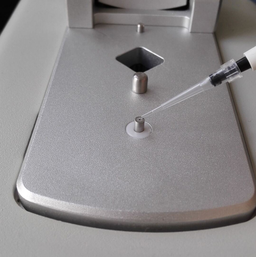
[Name] Medical Cold Patch
[Package Dimension] 10cm×12cm 2pieces/box
The pain relief patch is composed of three layers, namely, backing lining, middle gel and protective film. It is free from pharmacological, immunological or metabolic ingredients.
[Scope of Application] For cold physiotherapy, closed soft tissue only.
[Indications]
The patches give fast acting pain relief for strains, sprains, cramp, bruises, swollen areas or joint stiffness.
[How To Use a Patch]
Tear off the packaging bag and remove the patch. Remove the protective film and apply the patch on the skin. One piece, one time. The curing effect of each piece can last for 8-12 hours.
[Attention]
Do not apply the patch on the problematic skin, such as wounds, eczema, dermatitis,or in the eyes. People allergic to herbs and the pregnant are advised not to use the medication. If swelling or irritation occurs, please stop using and if any of these effects persist or worsen.notify your doctor or pharmacist promptly. Children using the patch must be supervised by adults.
[Storage Conditions]
Store below 30c in a dry place away from heat and direct sunlight.