Fluorescent Quantitative Pcr,Hiv Nucleic Acid Detection,Nucleic Acid Pcr Machine,Polymerase Chain Reaction Testing Jiangsu Dinai Bioengineering Co.,ltd. , https://www.dinaipcr.com
Bright field microscopy is a familiar method of microscopy. It is widely used in pathology and examination to observe stained sections. All microscopes can perform this function.
Bright field of vision 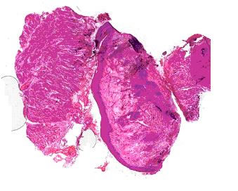
two. Dark field view (Dark field DF)
The dark field is actually a dark field illumination. Its characteristics are different from the bright field of view, and the light of the illumination is not directly observed, but the light reflected or diffracted by the object to be inspected is observed. Therefore, the field of view becomes a dark background, while the object being inspected presents a bright image.
The principle of the dark field is based on the optical phenomenon of Ding Dao. When the fine dust passes through the strong light, the human eye cannot observe it. This is caused by the glare of the strong light. If you illuminate it, the particles appear to increase in volume and are visible to the human eye due to the reflection of light.
The special accessory required for m..m dark field observation is a dark field condenser. It is characterized in that the light beam is not allowed to pass through the object to be inspected from bottom to top, but the light is redirected to the object to be inspected so that the illumination light does not directly enter the objective lens, and is formed by the surface reflection or diffracted light of the object to be inspected. Bright image. The resolution of dark field observation is much higher than that of bright field observation, up to 0.02-0.004
Dark field 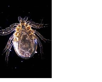
three. Phase contrast PH
In the development of optical microscopy, the successful invention of phase contrast microscopy is an important achievement in modern microscope technology. We know that the human eye can only distinguish the wavelength (color) and amplitude (brightness) of the light wave. For the colorless and bright biological specimen, when the light passes, the wavelength and amplitude do not change much, and it is difficult to observe the specimen when viewed in the bright field. .
The phase contrast microscope uses the difference between the optical paths of the object to be examined, that is, the interference phenomenon of the light is effectively utilized, and the phase difference that is indistinguishable to the human eye is changed into a resolvable amplitude difference, even if it is a colorless and transparent substance. Become clearly visible. This greatly facilitates the observation of living cells, so phase contrast microscopy is widely used in inverted microscopes.
Phase difference picture 
The basic principle of the phase contrast microscope is to change the optical path difference of visible light transmitted through the specimen into an amplitude difference, thereby improving the contrast between various structures and making various structures clearly visible. After the light passes through the specimen, it refracts, deviates from the original optical path, and is delayed by 1/4λ (wavelength). If it increases or decreases by 1/4λ, the optical path difference becomes 1/2λ, and the two beams of light combine with the rear interference. Strengthen, increase or decrease the amplitude, and increase the contrast. In terms of construction, phase contrast microscopy has two special features different from ordinary optical microscopy:
1. The annular diaphragm is located between the light source and the concentrator to form a hollow cone of light that passes through the concentrator and focus on the specimen.
2. The phase plate is a phase plate coated with magnesium fluoride in the objective lens to delay the phase of direct or diffracted light by 1/4λ. Divided into two types:
1. Phase A plate: The direct light is delayed by 1/4λ. After the two sets of light waves are combined, the light waves are added together, the amplitude is increased, and the specimen structure is brighter than the surrounding medium, forming a bright contrast (or negative contrast).
2. Phase B plate: delays the diffracted light by 1/4λ. After the two sets of rays are combined, the light wave is subtracted, the amplitude becomes smaller, and a dark contrast (or positive contrast) is formed. The structure is darker than the surrounding medium. 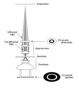
four. Differential interference contrast DIC
Differential interference microscopy appeared in the 1960s. It not only can observe colorless and transparent objects, but also the image shows a three-dimensional sense of relief and has some advantages that phase contrast microscopy can not achieve. lifelike.
principle;
Differential interference microscopy is the use of a special Wollaston prism to decompose the beam. The vibration directions of the split beams are perpendicular to each other and the intensity is equal. The beams pass through the object to be inspected at two points close to each other, and the phases are slightly different. Since the splitting distance of the two beams is extremely small, no ghost phenomenon occurs, and the image exhibits a three-dimensional three-dimensional feeling.
Differential interference picture 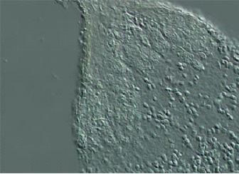
The physical principle of a DIC microscope is completely different from that of a phase contrast microscope, and the technical design is much more complicated. DIC uses polarized light and has four special optical components: a polarizer, a DIC prism, a DIC glider, and an analyzer. The polarizer is mounted directly in front of the concentrating system to linearly polarize the light. In the concentrator, a åŒmass prism, a DIC prism, is installed, which splits a beam of light into two beams of light (x and y) having different polarization directions, which are at a small angle. The concentrator adjusts the two beams into a direction parallel to the optical axis of the microscope. The first two beams have the same phase, and after passing through the adjacent areas of the specimen, the optical path difference between the two beams is caused by the difference in thickness and refractive index of the specimen. A second masculine prism, the DIC glider, is mounted at the back focal plane of the objective lens, which combines the two beams into one bundle.
At this time, the planes of polarization (x and y) of the two beams still exist. The last beam passes through a second polarizing device, the analyzer. The analyzer is at right angles to the direction of the polarizer before the beam forms the DIC image of the eyepiece. The analyzer combines two vertical beams of light into two beams of the same plane of polarization, thereby interfering with the two. The optical path difference between the x and y waves determines how much light is transmitted. When the optical path difference is 0, no light passes through the analyzer; when the optical path difference is equal to half of the wavelength, the passing light reaches a maximum value. So on a gray background, the specimen structure shows a bright and dark difference. In order to achieve the best contrast of the image, the optical path difference can be changed by adjusting the vertical fine adjustment of the DIC slider, and the optical path difference can change the brightness of the image. Adjusting the DIC glider can make the specimen's fine structure show a positive or negative projection image, usually one side is bright and the other side is dark, which causes the artificial three-dimensional sense of the specimen, similar to the principle of relief differential interference on marble. Figure 
5. Polarizing microscope POL
Characteristics of Polarized Microscopy Polarized light microscopy is a microscope that identifies the optical properties of a subtle structure. Any substance with birefringence can be clearly distinguished under a polarizing microscope. Of course, these substances can also be observed by dyeing, but some are impossible, and a polarizing microscope must be used.
The characteristic of polarized light microscopy is to change the ordinary to polarized light for microscopic examination to identify whether a substance is monorefracted (for each other) or birefringent (anisotropic).
Birefringence is a fundamental property of crystals. Therefore, polarized light microscopes are widely used in the fields of minerals, chemistry, and the like. It is also used in biology and botany.
6. Relief phase contrast microscope (RC HMC)
In 1975, Dr. Robert Hoffman invented
In 2002, the patent expired, and various microscope manufacturers have introduced the principle of RC technology products named in their own name. The oblique light is irradiated onto the specimen to produce refraction and diffraction. The light passes through the objective optical density gradient adjuster to produce different shadows, so that the surface of the transparent specimen is generated. Difference in brightness and darkness, increased observation contrast characteristics to improve the visibility and contrast of unstained specimens;
The image shows a shadow or an approximate three-dimensional structure without halos;
Detectable birefringent materials (rock slices, crystals, bones);
It can detect cells, organs and tissues in culture dishes such as glass and plastic;
The working distance of the condenser can be designed longer;
RC objectives can also be used for brightfield, darkfield and fluorescence observations
Seven: Fluorescence Microscopy FL
Fluoroscopy is the use of short-wavelength light to illuminate an object that has been stained with fluorescein, which is excited to produce long-wavelength fluorescence, which is then observed.
Fluorescent image 
advantage:
? High detection ability (magnification)
? Small stimulation to cells (can be stained in vivo)
? Can be used for multiple dyeing purposes:
? Observation of object structure - fluorescein
? The presence or absence of fluorescence, color comparison, material discrimination - antibody fluorescence, etc.
Fluorescence measurement for qualitative and quantitative analysis of fluorescence 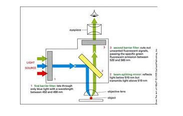
one. Bright field BF